HOW CANCER DEVELOPS
A healthy human body is composed of 30 trillion cells, most of which are in constant turnover as cells die and others reproduce to replace them in an orderly fashion. Healthy cells of the skin, hair, lining of the stomach, and blood, for example, regularly reproduce by dividing to form two daughter cells (see Mitosis). This cell division cycle proceeds under the regulation of the body’s intricately tuned control system. Among other functions, this control system ensures that cells only divide when needed, so that organs and tissues maintain their correct shape and size. Should this system fail, a variety of backup safety mechanisms prevent the cell from dividing uncontrollably. In order for a cell to become cancerous, every one of these safety mechanisms must fail.
Cancer begins in genes, bits of biochemical instructions composed of individual segments of the long, coiled molecule deoxyribonucleic acid (DNA). Genes contain the instructions to make proteins, molecular laborers that serve as building blocks of cells, control chemical reactions, or transport materials to and from cells. The proteins produced in a human cell determine the function of each cell, and ultimately, the function of the entire body.
In a cancerous cell, permanent gene alterations, or mutations, cause the cell to malfunction. For a cell to become cancerous, usually three to seven different mutations must occur in a single cell. These genetic mutations may take many years to accumulate, but the convergence of mutations enables the cell to become cancerous

DNA and Cancer
Cancer begins in the genes, segments of the long, coiled molecule known as deoxyribonucleic acid (DNA). Genes govern the body’s development and specific characteristics by providing critical instructions that trigger the production of proteins within the body. In cancer, certain genes fail to perform their jobs correctly. This computer-generated model shows two strands of deoxyribonucleic acid (DNA) and its double-helical structure.
Ken Eward/Photo Researchers, Inc.
While each human cell performs its own specialized function, it also exerts influence on the cells around it. Cells communicate with one another via receptors, protein molecules on the cell surface. A cell releases chemical messages, which fit into the surface receptors of cells nearby, much as a key fits into a lock. A cell may instruct other cells in its neighborhood to divide, for example, by releasing a growth-promoting signal, or growth factor. The growth factor binds to receptors on adjacent cells, activating a message within each individual cell. This message travels to the nucleus, where a cell’s genes are located.
Proto-Oncogenes Become Oncogenes
Nucleus of a Cell

The nucleus, present in eukaryotic cells, is a discrete structure containing chromosomes, which hold the genetic information for the cell. Separated from the cytoplasm of the cell by a double-layered membrane called the nuclear envelope, the nucleus contains a cellular material called nucleoplasm. Nuclear pores, present around the circumference of the nuclear membrane, allow the exchange of cellular materials between the nucleoplasm and the cytoplasm.
Don W. Fawcett/Science Source/Photo Researchers, Inc.
When the growth factor message reaches the cell nucleus, it activates genes called proto-oncogenes. These genes produce proteins that stimulate the cell to divide. In cancerous cells, mutations in proto-oncogenes cause these genes to malfunction. When a proto-oncogene mutates, it becomes an oncogene—a gene that instructs the cell to grow and divide repeatedly without stimulation from neighboring cells. Some oncogenes overproduce growth factors, causing the cell to divide too often. Other oncogenes stimulate the cell to reproduce even when no growth factor is present. Cancer researchers have identified about 100 different types of proto-oncogenes and their cancer-causing oncogene counterparts.
Tumor Suppressor Genes Stop Working
When runaway cell division occurs, it does not necessarily lead to cancer. Neighboring cells respond by excreting a growth inhibitor. This chemical binds to receptors in the malfunctioning cell, sending a signal to the nucleus that activates tumor suppressor genes. Tumor suppressor genes are like brakes for cell growth. When activated, these genes halt the cell cycle, preventing further cell division.
But if tumor suppressor genes malfunction due to mutations, the rapidly dividing cell ignores messages from its neighbors telling it to stop dividing. Malfunctioning tumor suppressor genes are not enough to cause cancer—the cell still must overcome a host of other safety mechanisms before it can cause truly significant damage.
Cell Cycle Clock Malfunctions
The cell nucleus contains a collection of interacting proteins that control cell division. Sometimes called the cell cycle clock, this group of proteins interprets incoming messages at several checkpoints in the cell division cycle. At these checkpoints, the clock evaluates the health of the cell. If conditions are right, the clock activates certain proto-oncogenes, which produce proteins that trigger the cell to enter the next stage of the cell cycle. If conditions are not right, certain tumor suppressor genes produce proteins that prevent the cell from proceeding with cell division.
If the cell cycle clock detects DNA damage in a cell, a tumor suppressor gene called p53 prevents the cell from reproducing until the damage is repaired. If the cell is unable to repair the DNA damage, p53 instructs the cell to undergo programmed cell death, or apoptosis, putting a stop to runaway cell division before it starts. Programmed cell death is a normal part of cell life and is tightly controlled by many genes, primarily p53.
In a cancerous cell, one or more mutations prevent these genes from doing their jobs. When mutated, p53 allows a cell to continue to divide, even with damaged DNA. This can lead to additional mutations in proto-oncogenes or tumor suppressor genes. In some cases, mutations occur in genes that produce proteins to repair damaged DNA. Such mutations can lead to yet other mutations because the faulty DNA cannot duplicate properly during cell division.
Cells Achieve Immortality
A normal cell has a life span of about 40 cell divisions. This life span is controlled in part by telomeres, protective segments at the ends of the cell’s DNA. Telomeres shorten with each cell division until they can no longer protect the DNA. At this point cell division severely damages the DNA, ultimately killing the cell. This normal process ensures that older cells, which may have accumulated mutations, no longer reproduce. Cancer cells escape this protective mechanism by producing a protein called telomerase. Telomerase extends the length of telomeres indefinitely, rendering the cells immortal and capable of never-ending cell division.
Cells Break Free and Spread
Evading the many obstacles that guard against runaway cell division is still not enough for cancer to develop. A malfunctioning cell must also skirt a number of safety mechanisms designed to prevent cells from growing where they are not supposed to in the body.
Normal cells adhere to each other and to a fibrous meshwork called an extracellular matrix. This matrix exists throughout all tissues and provides the structural support on which cells grow and form organs and other complex tissues. While a normal cell will often die if it cannot adhere to an extracellular matrix, cancer cells survive without this matrix.

Abnormal Cells and Cancer
Cancerous cells usually become much different from the tissue from which they arise. The ovarian tumor pictured here bears no resemblance to the normal tissue of the ovary.
Manfred Kage/Oxford Scientific Films
A tumor is a mass of cells not dependent upon an extracellular matrix. These cells can grow on top of each other, creating a mass of abnormal cells. Often a tumor develops its own network of tiny blood vessels to supply itself with nutrient-rich blood, a process called angiogenesis.
There are two general types of tumors. Benign tumors do not invade other tissues and are limited to one site, making surgical removal possible and the odds for a full recovery excellent. Some benign tumors are quite harmless and are not surgically removed unless they are unsightly or uncomfortable. For example, warts are benign tumors of the outer layer of the skin. Although they are usually not dangerous, warts may cause discomfort. Other benign tumors are thought to be precursors to cancerous, or malignant, tumors.
There are two general types of tumors. Benign tumors do not invade other tissues and are limited to one site, making surgical removal possible and the odds for a full recovery excellent. Some benign tumors are quite harmless and are not surgically removed unless they are unsightly or uncomfortable. For example, warts are benign tumors of the outer layer of the skin. Although they are usually not dangerous, warts may cause discomfort. Other benign tumors are thought to be precursors to cancerous, or malignant, tumors.
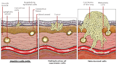
The Development and Spread of Tumors
Lung cancer begins when epithelial cells lining the respiratory tract start to reproduce in an uncontrolled fashion. These cells invade surrounding tissue, forming a mass called a tumor and, when hardened, a carcinoma. Cancerous cells may penetrate blood and lymph vessels, to be carried through the body until they reach a juncture through which they cannot pass. At this point, they lodge and new tumors form. Metastasis, the spreading of cancer from its original location to other parts of the body, is the disease’s most destructive characteristic
Tumors are malignant only if they can invade other parts of the body. Malignant tumors extend into neighboring tissue or travel to distant sites, forming secondary growths known as metastases. To metastasize, tumor cells break through a nearby blood vessel to enter the circulatory system or through a lymphatic vessel wall to enter the lymphatic system. Most metastases occur in organs that are the next site downstream in the circulatory system or the lymphatic system and contain a network of capillaries, or small blood vessels. For example, cancer of the large intestine often travels through the bloodstream to the liver, the organ immediately downstream from the intestines. In the lymphatic system, tumor cells can spread to surrounding lymph nodes, or lymph glands. Normally, lymph nodes filter out and destroy infectious materials circulating in the lymphatic system.
The unique receptors on the surface of a cell may also play a role in where tumors metastasize. Specialized molecules on a cell’s surface identify where in the body the cell belongs. Similar cells adhere to one another when their surface receptors are compatible. Most often cells from different tissues and organs have incompatible surface receptors. However, some tissue types share similar surface receptors, enabling cancerous cells to move between them and proliferate. Prostate cells and bone cells, for example, have similar surface receptors. This gives prostate cancer cells a natural affinity for bone tissue, where they can settle to form a new tumor.
Many cancers shed cells into the bloodstream early in their growth. Most of these cells die in the bloodstream, but some lodge against the surface of the blood vessel walls, eventually breaking through them and into adjacent tissue. In some cases, these cells survive and grow into a tumor. Others may divide only a few times, forming a small nest of cells that remain dormant as a micrometastasis. They may remain dormant for many years, only to grow again for reasons not yet known.
